Body Systems
Activity Overview:
You will identify the major components of each equine body system.
Directions:
1. Review each of the body system diagrams below.
2. Label the following items within each of the body systems provided. Do NOT use any reference materials to assist in the labeling process.
· For the muscular system, your instructor will determine the number of muscles you will need to identify.
3. Once completed, turn in your activity according to your instructor’s directions.
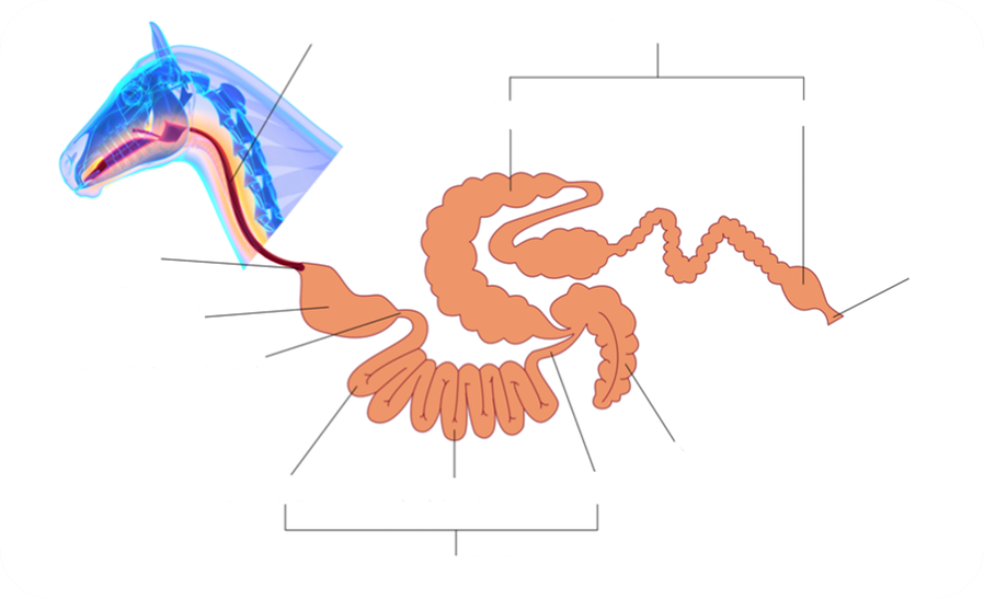
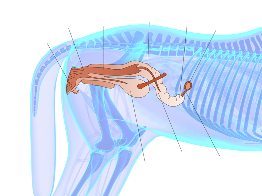
Digestive System
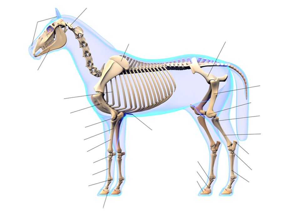
Skeletal System
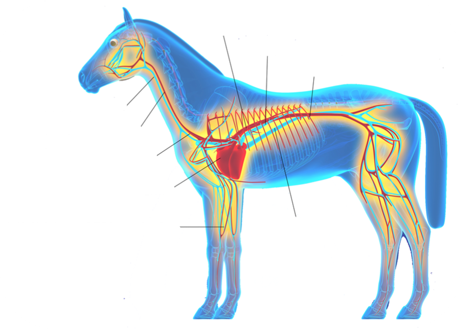
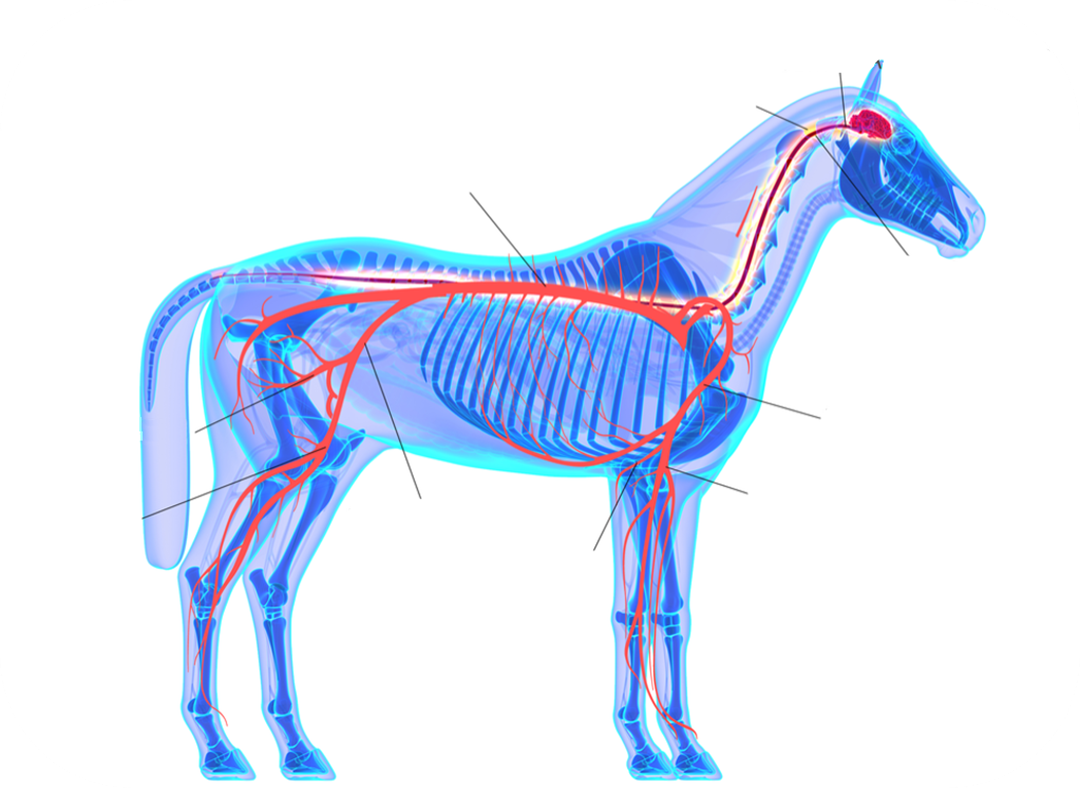
Circulatory System
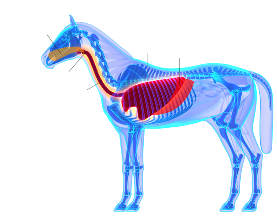
Respiratory System
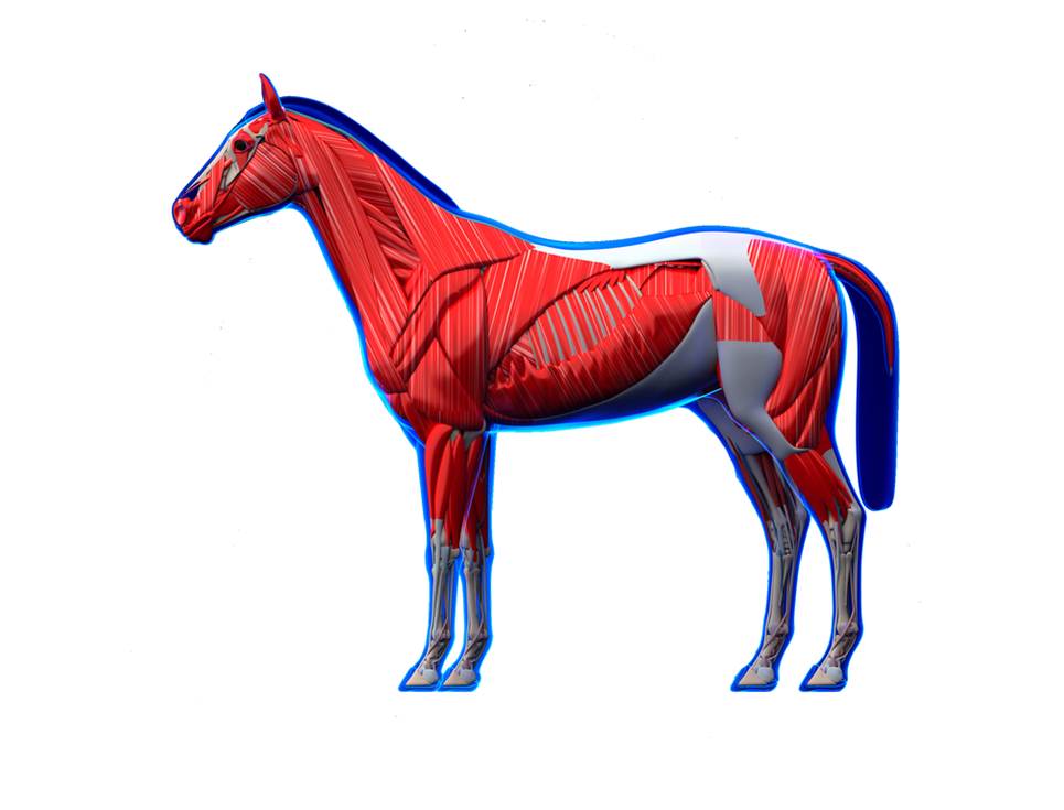
Muscular System
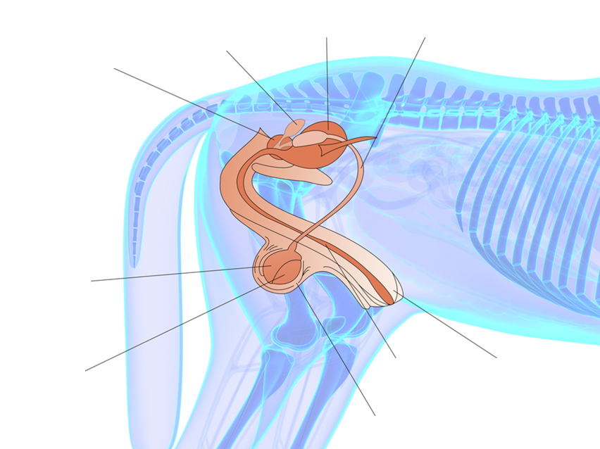
Reproductive System
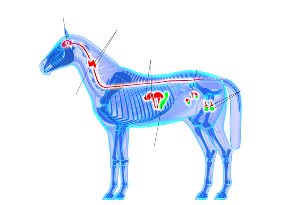
Endocrine System
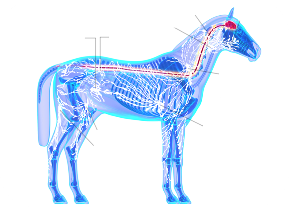
Immune System
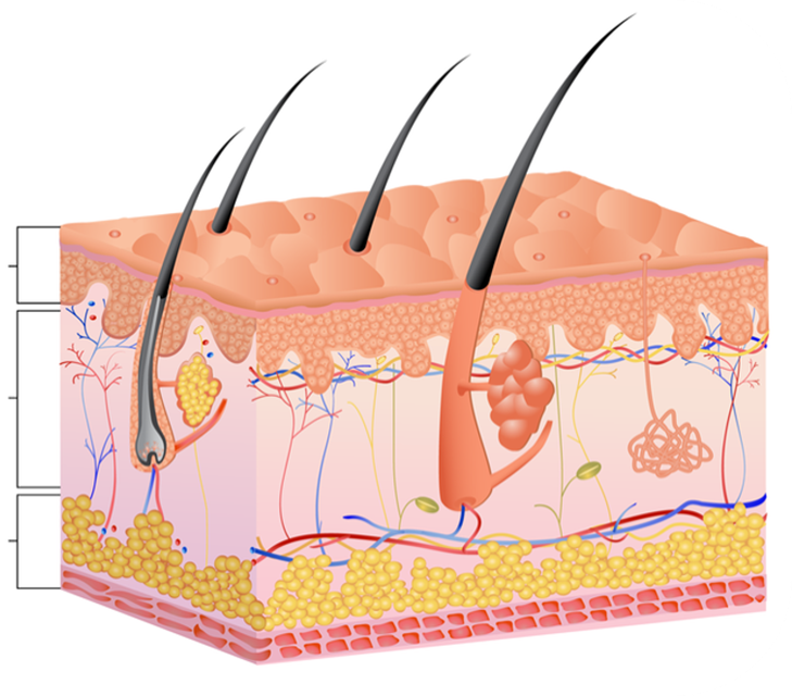
Integumentary System
Nervous System
![]()